Why did we create the X-ray Marker Parker™?
|
Radiologic technologists and other imaging professionals around the world utilize X-ray markers, also known as lead identification markers, to annotate medical images. X-ray markers help technologists identify which anatomical side of the body is being imaged, as well as other relevant information such as the time, date, patient position, etc. These markers are usually made of radiopaque lead letters, making the markers appear on an x-ray image, and sealed with an epoxy. Often, markers are relatively small, and may be held in the palm of a technologist’s hand. X-ray markers are commonly rectangular in shape with minimal thickness.
|
|
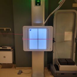
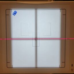
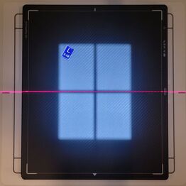
Generally, an X-ray marker may be attached directly to an imaging plate (detector, receptor, etc.), on a wall or table “bucky”, or on top of a patient. Regardless of the attaching surface, a marker may be placed in the area or field of view in which radiation passes through, also referred to as a “light field”, causing the marker to show up on an X-ray image. X-ray radiation passes through the patient and the X-ray marker and hits an imaging plate. The imaging plate then digitally processes an image, showing an anatomical artifact (from the patient) and a lead artifact (from the marker).
|
|
To attach these identification markers, an imaging technologist often utilizes tape, such as Transpore clear tape, or other double-sided adhesives to attach the marker to the imaging plate or another attaching surface. Generally, personal markers are carried by each technologist throughout a healthcare facility from patient to patient. The two most common personal markers are an “L” for left and an “R” for right, both of which may be annotated with the technologist’s initials. Most technologists attach their personal markers to their ID badge or a separate sheet of plastic with double-sided tape. Then the ID badge or plastic are often stored on a retractable reel or in scrub pockets.
|
This presents numerous problems. For one, when a technologist is working with a patient, touching their ID badge to grab their identification markers spreads germs and other bacteria between the patient and the ID badge. The same is true when a technologist reaches into a scrub pocket to grab their markers. In a hospital setting, ID badges are typically used for opening locked doors and logging into computers throughout the healthcare setting. Thus, when a technologist touches their ID badge during an exam, the risk of bacteria spreading everywhere the ID badge is used increases.
Secondly, removing and pulling a marker off a thin sheet of plastic, such as an ID badge, can be difficult. This can result in the wear and tear on an ID badge and the markers themselves. Furthermore, if a technologist attempts to avoid this issue by storing the markers without attaching them to plastic or another object, the double-sided tape on the back of the markers will remain exposed and accumulate dirt, lint, and other microorganisms.


